
📥 Instant Direct Digital Download
🕒 24/7 Support Available
💳 Payments Are Secure and Encrypted
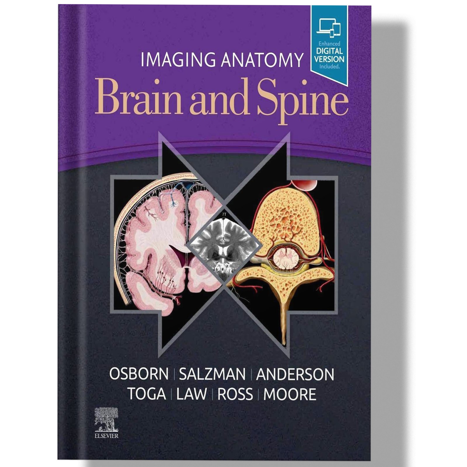
Imaging Anatomy Brain and Spine 1st Edition
-83% OffOriginal price was: $179.99.$29.99Current price is: $29.99.
Frequently Bought Together!

Original price was: $215.99.$29.99Current price is: $29.99.

Original price was: $152.99.$29.99Current price is: $29.99.
Related products
-
 -56% Off Original price was: $67.99.$29.99Current price is: $29.99.
📥 Instant Direct Digital Download
🕒 24/7 Support Available
💳 Payments Are Secure and Encrypted -
 -92% Off Original price was: $395.99.$29.99Current price is: $29.99.
📥 Instant Direct Digital Download
🕒 24/7 Support Available
💳 Payments Are Secure and Encrypted -
 -90% Off Original price was: $287.99.$29.99Current price is: $29.99.
📥 Instant Direct Digital Download
🕒 24/7 Support Available
💳 Payments Are Secure and Encrypted -
 -71% Off Original price was: $103.99.$29.99Current price is: $29.99.
📥 Instant Direct Digital Download
🕒 24/7 Support Available
💳 Payments Are Secure and Encrypted -
 -99% Off Original price was: $2,100.99.$29.99Current price is: $29.99.
📥 Instant Direct Digital Download
🕒 24/7 Support Available
💳 Payments Are Secure and Encrypted -
 -77% Off Original price was: $130.99.$29.99Current price is: $29.99.
📥 Instant Direct Digital Download
🕒 24/7 Support Available
💳 Payments Are Secure and Encrypted -
 -94% Off Original price was: $513.99.$29.99Current price is: $29.99.
📥 Instant Direct Digital Download
🕒 24/7 Support Available
💳 Payments Are Secure and Encrypted -
 -82% Off Original price was: $170.99.$29.99Current price is: $29.99.
📥 Instant Direct Digital Download
🕒 24/7 Support Available
💳 Payments Are Secure and Encrypted -
 -91% Off Original price was: $336.99.$29.99Current price is: $29.99.
📥 Instant Direct Digital Download
🕒 24/7 Support Available
💳 Payments Are Secure and Encrypted -
 -71% Off Original price was: $104.99.$29.99Current price is: $29.99.
📥 Instant Direct Digital Download
🕒 24/7 Support Available
💳 Payments Are Secure and Encrypted -
 -78% Off Original price was: $134.99.$29.99Current price is: $29.99.
📥 Instant Direct Digital Download
🕒 24/7 Support Available
💳 Payments Are Secure and Encrypted -
 -64% Off Original price was: $83.99.$29.99Current price is: $29.99.
📥 Instant Direct Digital Download
🕒 24/7 Support Available
💳 Payments Are Secure and Encrypted
Reviews
There are no reviews yet.